Kidney Stones: Types, Causes & Complete Treatment by UrologyMax
Nephrolithiasis — kidney stones — is one of the most prevalent and painful conditions in all of urology, affecting approximately 1 in 11 Americans over their lifetime, with a lifetime recurrence risk exceeding 50% in untreated patients. The Mid-Atlantic region, including Washington, D.C., Maryland, and Virginia, falls within the geographic "stone belt" — an area of heightened stone incidence attributable to a combination of climatic heat, regional dietary patterns, and relatively hard municipal water supplies. Modern stone management has been transformed by minimally invasive technology: while open surgical stone removal was standard as recently as the 1970s, virtually all stones that fail to pass spontaneously today are addressed with outpatient, minimally invasive procedures that require no incision and allow same-day or next-day return to normal activity.
If a stone can be seen on a plain x-ray, it is a calcium stone. These stones cannot be dissolved, and must either pass or be treated. On the contrary, uric acid stones comprise perhaps 10% of all stones, which if not coated with calcium, can be dissolved by raising the pH of the urine.
Stone composition varies and has direct implications for treatment and prevention. Calcium oxalate stones — the most prevalent type, accounting for approximately 75% of all cases — are radiopaque and visible on plain X-ray, but cannot be dissolved pharmacologically and must either pass or be removed procedurally. Calcium phosphate stones (approximately 10% of cases) behave similarly. Uric acid stones (roughly 10% of cases) are radiolucent — invisible on plain X-ray but detectable on CT — and offer an important therapeutic distinction: they can be dissolved over 4–8 weeks with oral urine-alkalinizing agents such as potassium citrate, provided they are not causing acute obstruction. Struvite stones form exclusively in the setting of urease-producing bacterial infections (Proteus, Klebsiella), may grow to fill the entire renal collecting system as staghorn calculi, and require both stone removal and definitive eradication of the causative organism.
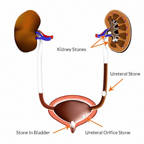
Kidney stones come to medical attention through three principal routes. Most dramatically, a stone enters the ureter and causes the sudden, severe onset of renal colic — flank pain radiating to the groin and inner thigh, often accompanied by nausea, vomiting, and visible or microscopic blood in the urine. Alternatively, a stone may be discovered incidentally on imaging performed for an unrelated reason — a CT scan ordered for abdominal pain, a renal ultrasound obtained during a physical examination, or a chest CT that captures the upper abdomen. The third presentation is hematuria workup: unexplained blood in the urine prompts radiological evaluation that identifies an asymptomatic calculus. Each pathway carries distinct management implications and treatment urgency.
Management of a newly diagnosed stone depends primarily on stone size, its location in the urinary tract, the presence or absence of obstruction, and whether signs of infection are present. For stones measuring 1–4 mm detected in the ureter, conservative management is appropriate and effective: high fluid intake targeting urine output greater than 2 liters per day, alpha-blocker therapy with tamsulosin 0.4 mg daily to relax ureteral smooth muscle and facilitate passage, adequate analgesic management, and urination through a urine strainer to confirm passage. Stones in this size range pass spontaneously approximately 70–80% of the time. Patients who do not retrieve the stone are re-imaged at 4 weeks to confirm passage or identify persistent obstruction.
However, larger stones that are 5mm or larger are more likely to become lodged in the ureter and not pass on their own. This is when a patient may be given a choice of waiting longer to see if it will pass or choosing to have the stone treated.
Ureteral stones measuring 5 mm or larger carry progressively lower rates of spontaneous passage — approximately 50–60% for 5–7 mm stones and as low as 10–15% for stones exceeding 8 mm. When a stone fails to pass within 4–6 weeks, is associated with uncontrollable pain, fever, or leukocytosis suggesting concurrent infection, is obstructing a solitary functional kidney, or is causing persistent symptomatic obstruction, procedural intervention is clearly indicated. UrologyMax offers both Extracorporeal Shock Wave Lithotripsy (ESWL) — a completely non-invasive acoustic fragmentation technique performed without any incision — and Ureteroscopy with Holmium Laser Lithotripsy, which provides direct visual access to the stone and achieves stone-free rates exceeding 95% for ureteral stones of any size. Treatment selection is individualized based on stone location, size, radiographic characteristics, and patient anatomy.
ESWL was popularized in the late 1980’s and early 1990’s. Before its introduction, open surgery had to be used. ESWL is a procedure whereby the patient is anesthetized and a machine called a lithotripter is used to fire sound waves at the stone. The original ESWL machines involved placing the anesthesized patient into a water bath as a means of transmitting the sound waves to the patient. Now, several generations later, these machines are portable and much smaller and simpler. ESWL can be used for stones lodged in the ureter, but is more often used for stones in the kidney. ESWL is the most minimally invasive procedure for kidney stones, but its success rates at producing a stone free kidney likely top out at 90% or so. Sometimes a stone will be treated with more than one lithotripsy or in combination with ureteroscopy.
Ureteroscopy was introduced, and became a very common treatment in the early 2000’s. What truly allowed ureterscopy to develop was the emergence of the use of lasers in Urology, and the progress made in fiber-optic technology. Now, ureteroscopes are very thin, are rigid or flexible, thus allowing nearly all stones to be treated this way. Here, instead of an x-ray guiding sound waves as in ESWL, the Urologist uses a scope to reach the stone directly, sees the stone on a video monitor, and uses a laser beam as the energy source to destroy the stone under direct vision. Ureteroscopy will generally require a temporary ureteral stent, which some patients would prefer to avoid but with some exceptions are generally well tolerated. Ureteroscopy is the most common way to treat ureteral stones but can treat kidney stones as well. Success rates with ureteroscopy are slightly higher than with ESWL, but it is a somewhat more invasive procedure that will require stent placement and stent removal at a later date. Both ESWL and ureteroscopy are typically done in an outpatient surgical setting, and it must be understood that in both cases small stone fragments will still need to be passed. The stones do not simply disappear.
As stated above, a percutaneous nephrolithotomy is a much more rare procedure. In the hands of Dr. Grant, this will be offered in the setting of a very large stone, perhaps 3cm or greater. Here, a needle is placed through the back directly into the kidney. This tract is used to place a tube called a nephrostomy tube, and this access will be used in the operating room to allow the Urologist direct access into the kidney with a much larger scope. A nephrolithotomy carries with it more risk such as bleeding, and will require an overnight hospital stay, but at times is the only viable option for some stones.
All patients must know that perhaps the most serious complication after a stone procedure is infection.
All patients must know that perhaps the most serious complication after a stone procedure is infection. Fevers after a procedure could signal infection behind an obstruction, an emergency situation that could rapidly progress to a condition called sepsis which can be life threatening. Fevers or feeling very sick after a stone procedure is always an emergency, so please dial 911 or go to your nearest or favorite emergency room and ensure that Dr. Grant or Dr. Tobon are contacted to take part in your care. As a reminder, UroMax physicians have privileges at Reston Hospital Center, Holy Cross Hospital Silver Spring, and Holy Cross Hospital Germantown.
If you have been diagnosed with a kidney or ureteral stone, we would be happy to review your films/cd with you during a consultation in our office and make recommendations.
Related Pages


Frequently Asked Questions
How do I schedule an appointment at UrologyMax?
Schedule online or call 1-800-876-6299 or use our online booking. Same-day and telehealth appointments are available.
Does UrologyMax accept insurance?
UrologyMax works with most major insurance plans. Contact our billing team at 1-800-876-6299 to verify your coverage before your visit.
What should I bring to my first urology appointment?
Bring your insurance card, photo ID, medication list, prior test results, and a completed patient intake form available on our website.
Are telehealth consultations available?
Yes. UrologyMax offers telehealth consultations for many urological concerns, including ED prescriptions, follow-up visits, and second opinions.
What makes UrologyMax different from other urology practices?
UrologyMax combines advanced robotic surgery capabilities with discreet online ED medication ordering and a patient-first approach to comprehensive urological care.